Nerves
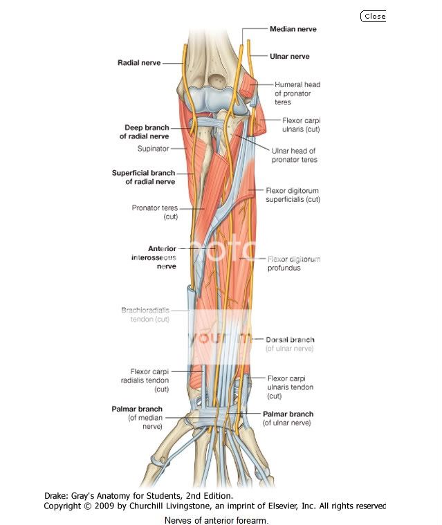
| Nerves in the anterior compartment of the forearm are the median and ulnar nerves, and the superficial branch of the radial nerve (Fig. 7.87). |
| Median nerve |
| The median nerve innervates the muscles in the anterior compartment of the forearm except for the flexor carpi ulnaris and the medial part of the flexor digitorum profundus (ring and little fingers). It leaves the cubital fossa by passing between the two heads of the pronator teres muscle and passing between the humero-ulnar and radial heads of the flexor digitorum superficialis muscle (Fig. 7.87). |
Most branches to the muscles in the superficial and intermediate layers of the forearm originate medially from the nerve just distal to the elbow joint.
|
| Ulnar nerve |
| The ulnar nerve passes through the forearm and into the hand, where most of its major branches occur. In the forearm, the ulnar nerve innervates only the flexor carpi ulnaris muscle and the medial part (ring and little fingers) of the flexor digitorum profundus muscle (Fig. 7.87). |
| Body_ID: P007518 |
| The ulnar artery is lateral to the ulnar nerve in the distal two-thirds of the forearm, and both the ulnar artery and nerve enter the hand by passing superficial to the flexor retinaculum and immediately lateral to the pisiform bone (Fig. 7.87). |
| Radial nerve |
- The deep branch is predominantly motor and passes between the two heads of the supinator muscle to access and supply muscles in the posterior compartment of the forearm.
- The superficial branch of the radial nerve is sensory. It passes down the anterolateral aspect of the forearm deep to the brachioradialis muscle and in association with the radial artery. Approximately two-thirds of the way down the forearm, the superficial branch of the radial nerve passes laterally and posteriorly around the radial side of the forearm deep to the tendon of the brachioradialis. The nerve continues into the hand where it innervates skin on the posterolateral surface.
From MOORE'S
Nerves of Forearm
Nerves of Forearm
The nerves of the forearm are the median, ulnar, and radial. The median nerve is the principal nerve of the anterior (flexorpronator) compartment of the forearm (Figs. 6.57B and 6.69A). Although the radial nerve appears in the cubital region, it soon enters the posterior (extensor-supinator) compartment of the forearm. Besides the cutaneous branches, there are only two nerves of the anterior aspect of the forearm: the median and ulnar nerves. The named nerves of the forearm are illustrated in Figure 6.69 and their origins and courses are described in Table 6.13. The following sections provide additional details and discuss unnamed branches.
MEDIAN NERVE IN FOREARM
The median nerve is the principal nerve of the anterior compartment of the forearm (Figs. 6.69A and 6.70; Table 6.13). It supplies muscular branches directly to the muscles of the superficial and intermediate layers of forearm flexors (except the FCU), and deep muscles (except for the medial [ulnar] half of the FDP) via its branch, the anterior interosseous nerve.
The median nerve has no branches in the arm other than small twigs to the brachial artery. Its major branch in the forearm is the anterior interosseous nerve (Fig. 6.69A, Table 6.13). In addition, the following unnamed branches of the median nerve arise in the forearm:
- Articular branches. These branches pass to the elbow joint as the median nerve passes it.
- Muscular branches. The nerve to the pronator teres usually arises at the elbow and enters the lateral border of the muscle. A broad bundle of nerves pierces the superficial flexor group of muscles and innervates the FCR, the palmaris longus, and the FDS.
- Anterior interosseous nerve. This branch runs distally on the interosseous membrane with the anterior interosseous branch of the ulnar artery. After supplying the deep forearm flexors (except the ulnar part of the FDP, which sends tendons to 4th and 5th fingers), it passes deep to and supplies the pronator quadratus, then ends by sending articular branches to the wrist joint.
- Palmar cutaneous branch of the median nerve. This branch arises in the forearm, just proximal to the flexor retinaculum, but is distributed to skin of the central part of the palm.
ULNAR NERVE IN FOREARM
Like the median nerve, the ulnar nerve does not give rise to branches during its passage through the arm. In the forearm it supplies only one and a half muscles, the FCU (as it enters the forearm by passing between its two heads of proximal attachment) and the ulnar part of the FDP, which sends tendons to the 4th and 5th digits (Fig. 6.69B, Table 6.13). The ulnar nerve and artery emerge from beneath the FCU tendon and become superficial just proximal to the wrist. They pass superficial to the flexor retinaculum and enter the hand by passing through a groove between the pisiform and the hook of the hamate.
A band of fibrous tissue from the flexor retinaculum bridges the groove to form the small ulnar canal (Guyon canal) (Fig. 6.70B). The branches of the ulnar nerve arising in the forearm include unnamed muscular and articular branches, and cutaneous branches that pass to the hand:
- Articular branches pass to the elbow joint while the nerve is between the olecranon and the medial epicondyle.
- Muscular branches supply the FCU and the medial half of the FDP.
- The palmar and dorsal cutaneous branches arise from the ulnar nerve in the forearm, but their sensory fibers are distributed to the skin of the hand.
RADIAL NERVE IN FOREARM
Unlike the medial and ulnar nerves, the radial nerve serves motor and sensory functions in both the arm and the forearm (but only sensory functions in the hand). However, its sensory and motor fibers are distributed in the forearm by two separate branches, the superficial (sensory or cutaneous) and deep radial/posterior interosseous nerve (motor) (Fig. 6.69C & D, Table 6.13). It divides into these terminal branches as it appears in the cubital fossa, anterior to the lateral epicondyle of the humerus, between the brachialis and the brachioradialis (Fig. 6.64). The two branches immediately part company, the deep branch winding laterally around the radius, piercing the supinator en route to the posterior compartment.
The posterior cutaneous nerve of the forearm arises from the radial nerve in the posterior compartment of the arm, as it runs along the radial groove of the humerus. Thus it reaches the forearm independent of the radial nerve, descending in the subcutaneous tissue of the posterior aspect of the forearm to the wrist, supplying the skin (Fig. 6.69D).
The superficial branch of the radial nerve is also a cutaneous nerve, but it gives rise to articular branches as well. It is distributed to skin on the dorsum of the hand and to a number of joints in the hand, branching soon after it emerges from the overlying brachioradialis and crosses the roof of the anatomical snuff box (Fig. 6.65).
The deep branch of the radial nerve, after it pierces the supinator, runs in the fascial plane between superficial and deep extensor muscles in close proximity to the posterior interosseous artery; it is usually referred to as the posterior interosseous nerve (Figs. 6.64 and 6.69C). It supplies motor innervation to all the muscles with fleshy bellies located entirely in the posterior compartment of the forearm (distal to the lateral epicondyle of the humerus).
LATERAL AND MEDIAL CUTANEOUS NERVES OF FOREARM
The lateral cutaneous nerve of the forearm (lateral antebrachial cutaneous nerve) is the continuation of the musculocutaneous nerve after its motor branches have all been given off to the muscles of the anterior compartment of the arm.
The medial cutaneous nerve of the forearm (medial antebrachial cutaneous nerve) is an independent branch of the medial cord of the brachial plexus. With the posterior cutaneous nerve of the forearm from the radial nerve, each supplying the area of skin indicated by its name, these three nerves provide all the cutaneous innervation of the forearm (Fig. 6.69D). There is no “anterior cutaneous nerve of the forearm.” (Memory device: This is similar to the brachial plexus, which has lateral, medial, and posterior cords but no anterior cord.)
Although the arteries, veins, and nerves of the forearm have been considered separately, it is important to place them into their anatomical context. Except for the superficial veins, which often course independently in the subcutaneous tissue, these neurovascular structures usually exist as components of neurovascular bundles. These bundles are composed of arteries, veins (in the limbs, usually in the form of accompanying veins), and nerves as well as lymphatic vessels, which are usually surrounded by a neurovascular sheath of varying density.


0 comments:
Post a Comment