Showing posts with label Palsy. Show all posts

Wednesday, August 5, 2009

Palsy (images)

Brachial plexus :
http://www.netterimages.com/image/4453.htm

Cutaneous innervation of the upper limb:
http://www.netterimages.com/image/4399.htm

Musculocutaneous nerve:
http://www.netterimages.com/image/4451.htm

Ulnar nerve:
http://www.netterimages.com/image/4611.htm

Radial nerve:
http://www.netterimages.com/image/4452.htm

Median nerve:
http://www.netterimages.com/image/4793.htm

Innervation of the Forearm

From GRAY'S ANATOMY

Nerves

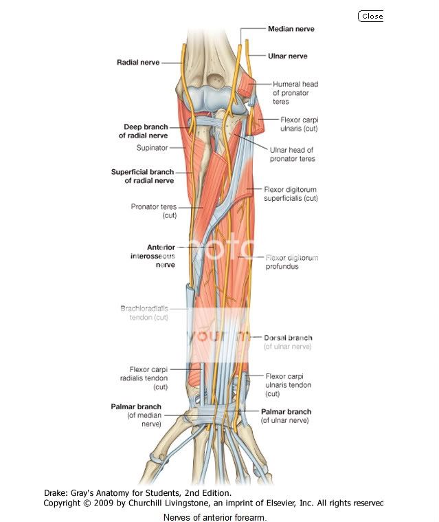
Nerves in the anterior compartment of the forearm are the median and ulnar nerves, and the superficial branch of the radial nerve (Fig. 7.87).

Median nerve

The median nerve innervates the muscles in the anterior compartment of the forearm except for the flexor carpi ulnaris and the medial part of the flexor digitorum profundus (ring and little fingers). It leaves the cubital fossa by passing between the two heads of the pronator teres muscle and passing between the humero-ulnar and radial heads of the flexor digitorum superficialis muscle (Fig. 7.87).

The median nerve continues a straight linear course distally down the forearm in the fascia on the deep surface of the flexor digitorum superficialis muscle. Just proximal to the wrist, it moves around the lateral side of the muscle and becomes more superficial in position, lying between the tendons of the palmaris longus and flexor carpi radialis muscles. It leaves the forearm and enters the palm of the hand by passing through the carpal tunnel deep to the flexor retinaculum.

Most branches to the muscles in the superficial and intermediate layers of the forearm originate medially from the nerve just distal to the elbow joint.
  • The largest branch of the median nerve in the forearm is the anterior interosseous nerve, which originates between the two heads of the pronator teres, passes distally down the forearm with the anterior interosseous artery, innervates the muscles in the deep layer (the flexor pollicis longus, the lateral half of flexor digitorum profundus, and pronator quadratus) and terminates as articular branches to joints of the distal forearm and wrist.
  • A small palmar branch originates from the median nerve in the distal forearm immediately proximal to the flexor retinaculum (Fig. 7.87), passes superficially into the hand and innervates the skin over the base and central palm. This palmar branch is spared in carpal tunnel syndrome because it passes into the hand superficial to the flexor retinaculum of the wrist.

Ulnar nerve

The ulnar nerve passes through the forearm and into the hand, where most of its major branches occur. In the forearm, the ulnar nerve innervates only the flexor carpi ulnaris muscle and the medial part (ring and little fingers) of the flexor digitorum profundus muscle (Fig. 7.87).
Body_ID: P007518
The ulnar nerve enters the anterior compartment of the forearm by passing posteriorly around the medial epicondyle of the humerus and between the humeral and ulnar heads of the flexor carpi ulnaris muscle. After passing down the medial side of the forearm in the plane between the flexor carpi ulnaris and the flexor digitorum profundus muscles, it lies under the lateral lip of the tendon of the flexor carpi ulnaris proximal to the wrist.

The ulnar artery is lateral to the ulnar nerve in the distal two-thirds of the forearm, and both the ulnar artery and nerve enter the hand by passing superficial to the flexor retinaculum and immediately lateral to the pisiform bone (Fig. 7.87).

In the forearm the ulnar nerve gives rise to:
  • muscular branches to the flexor carpi ulnaris and to the medial half of the flexor digitorum profundus that arise soon after the ulnar nerve enters the forearm; and
  • two small cutaneous branches-the palmar branch originates in the middle of the forearm and passes into the hand to supply skin on the medial side of the palm; the larger dorsal branch originates from the ulnar nerve in the distal forearm and passes posteriorly deep to the tendon of the flexor carpi ulnaris and innervates skin on the posteromedial side of the back of the hand and most skin on the posterior surfaces of the medial one and one-half digits.

Radial nerve
The radial nerve bifurcates into deep and superficial branches under the margin of the brachioradialis muscle in the lateral border of the cubital fossa (Fig. 7.87).
  • The deep branch is predominantly motor and passes between the two heads of the supinator muscle to access and supply muscles in the posterior compartment of the forearm.
  • The superficial branch of the radial nerve is sensory. It passes down the anterolateral aspect of the forearm deep to the brachioradialis muscle and in association with the radial artery. Approximately two-thirds of the way down the forearm, the superficial branch of the radial nerve passes laterally and posteriorly around the radial side of the forearm deep to the tendon of the brachioradialis. The nerve continues into the hand where it innervates skin on the posterolateral surface.


From MOORE'S

Nerves of Forearm

The nerves of the forearm are the median, ulnar, and radial. The median nerve is the principal nerve of the anterior (flexorpronator) compartment of the forearm (Figs. 6.57B and 6.69A). Although the radial nerve appears in the cubital region, it soon enters the posterior (extensor-supinator) compartment of the forearm. Besides the cutaneous branches, there are only two nerves of the anterior aspect of the forearm: the median and ulnar nerves. The named nerves of the forearm are illustrated in Figure 6.69 and their origins and courses are described in Table 6.13. The following sections provide additional details and discuss unnamed branches.
MEDIAN NERVE IN FOREARM
The median nerve is the principal nerve of the anterior compartment of the forearm (Figs. 6.69A and 6.70; Table 6.13). It supplies muscular branches directly to the muscles of the superficial and intermediate layers of forearm flexors (except the FCU), and deep muscles (except for the medial [ulnar] half of the FDP) via its branch, the anterior interosseous nerve.
The median nerve has no branches in the arm other than small twigs to the brachial artery. Its major branch in the forearm is the anterior interosseous nerve (Fig. 6.69A, Table 6.13). In addition, the following unnamed branches of the median nerve arise in the forearm:
  • Articular branches. These branches pass to the elbow joint as the median nerve passes it.
  • Muscular branches. The nerve to the pronator teres usually arises at the elbow and enters the lateral border of the muscle. A broad bundle of nerves pierces the superficial flexor group of muscles and innervates the FCR, the palmaris longus, and the FDS.
  • Anterior interosseous nerve. This branch runs distally on the interosseous membrane with the anterior interosseous branch of the ulnar artery. After supplying the deep forearm flexors (except the ulnar part of the FDP, which sends tendons to 4th and 5th fingers), it passes deep to and supplies the pronator quadratus, then ends by sending articular branches to the wrist joint.
  • Palmar cutaneous branch of the median nerve. This branch arises in the forearm, just proximal to the flexor retinaculum, but is distributed to skin of the central part of the palm.
ULNAR NERVE IN FOREARM
Like the median nerve, the ulnar nerve does not give rise to branches during its passage through the arm. In the forearm it supplies only one and a half muscles, the FCU (as it enters the forearm by passing between its two heads of proximal attachment) and the ulnar part of the FDP, which sends tendons to the 4th and 5th digits (Fig. 6.69B, Table 6.13). The ulnar nerve and artery emerge from beneath the FCU tendon and become superficial just proximal to the wrist. They pass superficial to the flexor retinaculum and enter the hand by passing through a groove between the pisiform and the hook of the hamate.
A band of fibrous tissue from the flexor retinaculum bridges the groove to form the small ulnar canal (Guyon canal) (Fig. 6.70B). The branches of the ulnar nerve arising in the forearm include unnamed muscular and articular branches, and cutaneous branches that pass to the hand:
  • Articular branches pass to the elbow joint while the nerve is between the olecranon and the medial epicondyle.

  • Muscular branches supply the FCU and the medial half of the FDP.
  • The palmar and dorsal cutaneous branches arise from the ulnar nerve in the forearm, but their sensory fibers are distributed to the skin of the hand.

RADIAL NERVE IN FOREARM
Unlike the medial and ulnar nerves, the radial nerve serves motor and sensory functions in both the arm and the forearm (but only sensory functions in the hand). However, its sensory and motor fibers are distributed in the forearm by two separate branches, the superficial (sensory or cutaneous) and deep radial/posterior interosseous nerve (motor) (Fig. 6.69C & D, Table 6.13). It divides into these terminal branches as it appears in the cubital fossa, anterior to the lateral epicondyle of the humerus, between the brachialis and the brachioradialis (Fig. 6.64). The two branches immediately part company, the deep branch winding laterally around the radius, piercing the supinator en route to the posterior compartment.
The posterior cutaneous nerve of the forearm arises from the radial nerve in the posterior compartment of the arm, as it runs along the radial groove of the humerus. Thus it reaches the forearm independent of the radial nerve, descending in the subcutaneous tissue of the posterior aspect of the forearm to the wrist, supplying the skin (Fig. 6.69D).
The superficial branch of the radial nerve is also a cutaneous nerve, but it gives rise to articular branches as well. It is distributed to skin on the dorsum of the hand and to a number of joints in the hand, branching soon after it emerges from the overlying brachioradialis and crosses the roof of the anatomical snuff box (Fig. 6.65).
The deep branch of the radial nerve, after it pierces the supinator, runs in the fascial plane between superficial and deep extensor muscles in close proximity to the posterior interosseous artery; it is usually referred to as the posterior interosseous nerve (Figs. 6.64 and 6.69C). It supplies motor innervation to all the muscles with fleshy bellies located entirely in the posterior compartment of the forearm (distal to the lateral epicondyle of the humerus).

LATERAL AND MEDIAL CUTANEOUS NERVES OF FOREARM
The lateral cutaneous nerve of the forearm (lateral antebrachial cutaneous nerve) is the continuation of the musculocutaneous nerve after its motor branches have all been given off to the muscles of the anterior compartment of the arm.
The medial cutaneous nerve of the forearm (medial antebrachial cutaneous nerve) is an independent branch of the medial cord of the brachial plexus. With the posterior cutaneous nerve of the forearm from the radial nerve, each supplying the area of skin indicated by its name, these three nerves provide all the cutaneous innervation of the forearm (Fig. 6.69D). There is no “anterior cutaneous nerve of the forearm.” (Memory device: This is similar to the brachial plexus, which has lateral, medial, and posterior cords but no anterior cord.)
Although the arteries, veins, and nerves of the forearm have been considered separately, it is important to place them into their anatomical context. Except for the superficial veins, which often course independently in the subcutaneous tissue, these neurovascular structures usually exist as components of neurovascular bundles. These bundles are composed of arteries, veins (in the limbs, usually in the form of accompanying veins), and nerves as well as lymphatic vessels, which are usually surrounded by a neurovascular sheath of varying density.

Erb's Palsy

Erb's palsy is a common birth injury resulting in paralysis of the upper brachial plexus. It is usually caused by a forcible traction at the neck region during a difficult delivery resulting in stretching of one or both sides of the cervical nerve roots. The shoulder of the baby could get caught and stretched behind the pubic symphysis bone during the strain of childbirth. Once this happen,the brachial plexus can be compressed,stretched or torn. This leaves the child with differing degrees of paralysis to the shoulder, arm or hand.

http://www.dorsetrehab.com/pdf/Erb-s_Palsy.pdf


Is Erb's palsy avoidable?

It can be avoided by having good health care during pregnancy such as:

1.

Blood sugars of mothers with diabetes mellitus or gestational diabetes require vigilant monitoring. They also require good dietary teaching, and tight control of blood sugars through diet or medication administration throughout the pregnancy. We know that high blood sugars "overnourish" the baby and make it gain weight faster than normal, and larger babies are more likely to get stuck in the birth canal.
2.

One option may be to consider an elective cesarean delivery rather than vaginal delivery, but this decision should not be made lightly either, since it also involves risks.

Risk factors that are more likely to result in difficult deliveries and Erb's Palsy:

*

Mothers who have had a prior child with shoulder dystocia, regardless of whether the previous child had a brachial plexus injury
*

Mothers who have had their labor induced or speeded up with drugs like Cervadil, Pitocin, or Cytotec
*

Larger babies, predictably, are born to mother's with diabetes, or gestational diabetes, particularly if the blood sugars have not been carefully monitored and managed
*

Mothers with smaller or unusual shaped pelvises or pelvic openings
*

Larger babies (especially those weighing over 8 1/2 pounds at birth)
*

Prolonged labors
*

Precipitous deliveries
*

Breech position
*

Fetal malposition in the birth canal

Since Erb's palsy is caused by the baby's shoulders being stuck and caught in the pubic symphysis druing vaginal delivery...
there are several techniques and maneuvers to dislodge the stuck shoulder safely. A team of nurses and doctors with current knowledge and skill in the techniques for these deliveries are less likely to deliver an injured baby.

Below is an illustration of maneuvers that nurses and physicians should employ to dislodge the baby when it has gotten stuck. Research demonstrates that rehearsals or drills by the labor team reduce the risk of injuries to the fetus when a true emergency occurs.


http://www.erbspalsyonline.com/


Treatment of Erb's palsy


Primary Surgery

Infants with mild injuries who do not heal by 3 to 4 months of age, or those with more severe injuries (such as avulsions or ruptures), need surgery to improve or correct nerve function. This surgery is best performed by a highly skilled, experienced team and ideally should occur within three to six months after birth. After children turn 1 year old, nerve surgery may not be as successful.

Among the techniques include:
* Neurolysis — clearing scar tissue from the nerve
* Nerve graft — a nerve is transplanted from the infant's leg to reconnect the damaged nerve(s)
* Nerve transfer — sewing an adjacent, functioning nerve or part of a nerve into a nonfunctioning nerve in an attempt to restore function in a paralyzed muscle.

Secondary Surgery

When there is less than full recovery, other conditions sometimes develop involving neighboring joints of the arm. These conditions can result in muscle imbalance or shortening of the muscle (contractures). In these cases, other procedures can be performed when the child is older, typically between ages 2 and 10. Possible procedures include:

* Free muscle transfer
* Capsule release
* Tendon transfer
* Correction of the arm (osteotomy)
* Joint fusion

Free muscle transfer-
When an injury causes irreversible atrophy (weakening) of the arm muscles, a new muscle can be transplanted to restore function. The surgical team will move an expendable muscle (such as the gracilis muscle from the thigh) along with its nerve and blood supply, to "reanimate" (restore movement to) the elbow, wrist and hand. Muscle transfers can often stabilize the shoulder and allow lifting of the shoulder, flexing of the elbow and, in some cases, restored function and sensation in the hand.

Tendon transfer-
A procedure where a healthy tendon is taken and moved to replace the function of a diseased or inactive tendon.

Capsule release-
Arthroscopic capsular release is keyhole surgery involving the release of the tight capsule seen in 'frozen shoulder'.

Oateotomy-
A surgical operation whereby a bone is cut to shorten, lengthen, or change its alignment.

Athrodesis(joint fusion)-
A surgical procedure which fuses the bones that form a joint, essentially eliminating the joint. The procedure is commonly referred to as joint fusion.Surgeons implant pins, plates, screws, wires, or rods to position the bones together until they fuse. Bone grafts are sometimes needed if there is significant bone loss. If bone grafting is necessary, bone can be taken from another part of the body or obtained from a bone bank.


http://www.mayoclinic.org/brachial-plexus/erbs-palsy.html