Showing posts with label Anatomy. Show all posts

Monday, September 28, 2009

Anatomy Dissections

Remember those dissection videos I showed before the Raya holidays - Clickie.

There's also another page on neuroscience with some neuroanatomy - Clickie.

Neuroscience dissection is here - Clickie.

You can save the videos if you want to, but it involves a bit of work - which I'm too lazy to type out here. You can either Google on how to save Quicktime files, or just bug me about it irl (in real life).

Monday, September 14, 2009

Superficial Anatomy of Buttock

Activity 1: Bones
1.1
Ilium, ischium, pubis
1.1.1
Ischium
1.1.1.1
Posterior division of anterior rami.

1.2


1.2.1.1
Bone marrow harvesting
1.2.2.1
Tensor of fascia latae, sartorius
1.2.3.1
A contusion (bruise) on the pelvis caused by a direct blow to an iliac crest. It usually causes bleeding into the hip abductor muscles, which move legs sideways, away from the midline of the body. This bleeding into muscle tissue creates swelling and makes leg movement painful. The injury usually last from one to six weeks, depending on the damage.

1.2.7.1
Superior gemelli
1.2.8.1
Sacrotuberous ligament, sacrospinous ligament and ischial spine. The sacrotuberous ligament extends across the sciatic notch converting the notch into a foramen that is further subdivided by the sacrospinous ligament and ischial spine into the greater and lesser sciatic foramen.
1.2.9.1
It is the site where the body’s weight rests on.
1.2.9.2
Inferior part of buttock when thigh is flexed.
1.2.9.3
The sciatic nerve extends from a point midway between the greater trochanter and the ischial tuberosity down the middle of the posterior aspect of the thigh. Hamstrings, biceps femoris and semitendinosus which attach proximally to the ischial tuberosity may avulse.

1.2.12.1
Inguinal ligament and indirect muscle attachments.
1.2.12.2
Provides the landmark for palpation of femoral pulse. By placing the tip of the little finger (of the right hand when dealing with the right side) on the ASIS and the tip of the thumb on the pubic tubercle, the femoral pulse can be palpated with the midpalm just inferior to the midpoint of the inguinal ligament by pressing firmly.

1.3.1
Obturator nerve and obturator artery.
1.3.2
Inferior epigastric artery.
1.3.2.1
Could be involved in strangulated femoral hernia. Surgeons placing staples during endoscopic repair of both inguinal and femoral hernias must be careful.

Activity 2: Muscles & Fascia


2.1.1
Proximal: ASIS, anterior part of iliac crest
Distal : Iliotibial trac, which attaches to lateral condyle of tibia.
2.1.2
Gluteus maximus.
2.1.3
Superior gluteal nerve( L5,S1)

2.2.1
Trochanteric bursa , the ischial bursa and gluteofemoral bursa.

2.2.2
Separate the gluteus maximus from adjacent structures, reduce friction and permit free movement.

2.2.3
A type of friction bursitis resulting from excessive friction between the ischial bursa and ischial tuberosities.

2.2.3.1
Recurrent mcirotrauma resulting from repeated stress eg : cycling, rowing or other activities involving hip extension while seated.


2.3.1
Superior gluteal nerve
2.3.2
Abduct and medially rotate thigh: keep pelvis level when ipsilateral limb is weight-bearing and advance opposite side during swing phase.

Activity 3: Vessels
3.1
Superior and inferior gluteal arteries.
3.2
Internal iliac artery which is a branch of the common iliac artery
3.2.1


Activity 4: Nerves
4.1 Inferior gluteal nerve
4.1.1
Compression and ischemia in sedentary individuals.
4.1.2
Difficulty in rising from seated position and climbing stairs.

4.2
Injections into the buttock are safe only in the superolateral quadrant of the buttock or superior to a line extending from the PSIS to the superior border of the greater trochanter (approximating the superior border of the gluteus maximus). IM injections can also be given safely into the anterolateral part of the thigh, where the needle enters the tensor fasciae latae as it extends distally from the iliac crest and ASIS. The index finger is placed on the ASIS, and the fingers are spread posteriorly along the iliac crest until the tubercle of the crest is felt by the middle finger . An IM injection can be made safely in the triangular area between the fingers (just anterior to the proximal joint of the middle finger) because it is superior to the sciatic nerve.

Friday, September 4, 2009

Anatomy of the eye

The human eyeball, the organ responsible for the sense of sight, is a very complex structure. We use our vision in almost every activity, so the eye is one of the most important organs in the body.

How it works
Sight begins when light rays from an object enter the eye through the cornea, the clear front “window” of the eyeball. The cornea is actually responsible for about sixty percent of the eyeball’s light-ray-bending capability. The cornea’s refractive power bends the light rays in such a way that they pass freely through the pupil, the size-changing hole in the iris. The iris, the structure that gives the eye color, works like a shutter in a camera. It has the ability to enlarge and shrink, depending on how much light the environment is sending into the eye.

After passing through the iris, the light rays strike the eye’s crystalline lens. This clear, flexible structure works much like the lens in a camera – shortening and lengthening its width in order to focus light rays properly.
In a normal eye, after exiting the back of the lens, the light rays pass through the vitreous -- a clear, jelly-like substance that fills the globe of the eyeball. The vitreous humor helps the eye hold its spherical shape. Finally, the light rays land and come to a sharp focusing point on the retina.
Continuing with our “camera” analogy, the retina’s function is much like the film in a camera. It is responsible for capturing all of the light rays, processing them into light impulses through millions of tiny nerve endings, then sending these light impulses through over a million nerve fibers to the optic nerve.The optic nerve is sort of like an extension of the brain. It is a bundled cord of more than a million nerve fibers. The light impulses travel through this nerve fiber to the brain, where they are interpreted as an image.

Wednesday, August 19, 2009

Anatomy of the Ear

Sorry i cant seem to get the pictures in here is the link so you guys can read.. some of the terms used are a lil different so i put them in brackets in the article i posted:

http://www.webschoolsolutions.com/patts/systems/ear.htm

Here is another site......... Anatomy of the ear made FUN^_^:

http://www.wisc-online.com/objects/index_tj.asp?objID=AP1502

Anatomy of the Ear

Introduction
The ears are paired sensory organs comprising the auditory system, involved in the detection of sound, and the vestibular system, involved with maintaining body balance/ equilibrium. The ear divides anatomically and functionally into three regions: the external ear, the middle ear, and the inner ear. All three regions are involved in hearing. Only the inner ear functions in the vestibular system.
Anatomy of the Ear
The external ear (or pinna, the part you can see) serves to protect the tympanic membrane (eardrum), as well to collect and direct sound waves through the ear canal(external acoustic meatus) to the eardrum. About 1¼ inches long, the canal contains modified sweat glands that secrete cerumen, or earwax. Too much cerumen can block sound transmission.


The middle ear, separated from the external ear by the eardrum, is an air-filled cavity (tympanic cavity) carved out of the temporal bone. It connects to the throat/nasopharynx via the Eustachian tube(pharyngotympanic tube). This ear-throat connection makes the ear susceptible to infection (otitis media). The eustachian tube functions to equalize air pressure on both sides of the eardrum. Normally the walls of the tube are collapsed. Swallowing and chewing actions open the tube to allow air in or out, as needed for equalization. Equalizing air pressure ensures that the eardrum vibrates maximally when struck by sound waves.
Adjoining the eardrum are three linked, movable bones called "ossicles," which convert the sound waves striking the eardrum into mechanical vibrations. The smallest bones in the human body, the ossicles are named for their shape. The hammer (malleus) joins the inside of the eardrum. The anvil (incus), the middle bone, connects to the hammer and to the stirrup (stapes). The base of the stirrup, the footplate, fills the oval window which leads to the inner ear.
The inner ear consists of a maze of fluid-filled tubes, running through the temporal bone of the skull. The bony tubes, the bony labyrinth, are filled with a fluid called perilymph. Within this bony labyrinth is a second series of delicate cellular tubes, called the membranous labyrinth, filled with the fluid called endolymph. This membranous labyrinth contains the actual hearing cells, the hair cells of the organ of Corti(spiral organ). There are three major sections of the bony labyrinth:

The front portion is the snail-shaped cochlea, which functions in hearing.
The rear part, the semicircular canals, helps maintain balance.
Interconnecting the cochlea and the semicircular canals is the vestibule, containing the sense organs responsible for balance, the utricle and saccule.
The inner ear has two membrane-covered outlets into the air-filled middle ear - the oval window and the round window. The oval window sits immediately behind the stapes, the third middle ear bone, and begins vibrating when "struck" by the stapes. This sets the fluid of the inner ear sloshing back and forth. The round window serves as a pressure valve, bulging outward as fluid pressure rises in the inner ear. Nerve impulses generated in the inner ear travel along the vestibulocochlear nerve (cranial nerve VIII), which leads to the brain. This is actually two nerves, somewhat joined together, the cochlear nerve for hearing and the vestibular nerve for equilibrium.


How We Hear - The Auditory System

All sounds (music, voice, a mouse-click, etc.) send out vibrations, or sound waves. Sound waves do not travel in a vacuum, but rather require a medium for sound transmission, e.g. air or fluid. What actually travels are alternating successions of increased pressure in the medium, followed by decreased pressure. These vibrations occur at various frequencies, not all of which the human ear can hear. Only those frequencies ranging from 20 to 20,000 Hz (Hz = hertz = cycles/sec) can be perceived.

In hearing, air-borne sound waves funnel down through the ear canal and strike the eardrum, causing it to vibrate. The vibrations are passed to the small bones of the middle ear (ossicles), which form a system of interlinked mechanical levers: First, vibrations pass to the malleus (hammer), which pushes the incus (anvil), which pushes the stapes (stirrup). The base of the stapes rocks in and out against the oval window - this is the entrance for the vibrations. The stapes agitates the perilymph of the bony labyrinth. At this point, the vibrations become fluid-borne. The perilymph, in turn, transmits the vibrations to the endolymph of the membranous labyrinth and, thence, to the hair cells of the organ of Corti. It is the movement of these hair cells which convert the vibrations into nerve impulses. The round window dissipates the pressure generated by the fluid vibrations, thus serves as the release valve: It can push out or expand as needed. The nerve impulses travel over the cochlear nerve to the auditory cortex of the brain, which interprets the impulses as sound.

Tuesday, August 11, 2009

Axilla Practical

Anatomy Practical: Axilla
Activity 1: Axilla and Other Spaces
i) The axilla is the pyramidal space inferior to the glenohumeral joint and superior to the axillary fascia at the junction of the arm and thorax.
 The anterior axillary fold is the inferiormost part of the anterior wall that may be grasped between the fingers; it is formed by the pectoralis major, as it bridges from thoracic wall to humerus, and the overlying integument.
 The posterior axillary fold is the inferiormost part of the posterior wall that may be grasped. It extends farther inferiorly than the anterior wall and is formed by latissimus dorsi, teres major, and overlying integument.
ii)
• -superiorly:
o and ventrally: subscapularis
o and dorsally: teres minor
• inferiorly: teres major
• medially: long head of triceps
• laterally: humerus
-It transmits the axillary nerve, posterior circumflex humeral vein, posterior circumflex humeral artery.
iii) It has the following boundaries:
• the Teres major inferiorly
• the long head of the Triceps laterally
• For the superior border, some sources list the Teres minor, while others list the Subscapularis.
-Contents: the scapular circumflex artery and vein.
-It arises from the subscapular artery.
-It arises together with the thoracodorsal artery.
The latissimus dorsi.
Activity 2: Blood Supply
i) The axillary artery begins at the lateral border of the 1st rib as the continuation of the subclavian artery and ends at the inferior border of the teres major. It passes posterior to the pectoralis minor into the arm and becomes the brachial artery when it passes the inferior border of the teres major, at which point it usually has reached the humerus.
 The first part of the axillary artery is located between the lateral border of the 1st rib and the medial border of the pectoralis minor; it is enclosed in the axillary sheath.
The second part of the axillary artery lies posterior to pectoralis minor and has two branches.
The third part of the axillary artery extends from the lateral border of pectoralis minor to the inferior border of teres major.
 First part has 1 branch-superior thoracic artery
Second part( 2 brahcnes)-thoracoacromial and lateral thoracic arteries.
Third part( 3 branches)-subscapular artery( largest branch), anterior circumflex humeral and posterior circumflex humeral arteries.
 Subscapular artery receives blood through several anastomoses with the suprascapular artery, dorsal scapular artery, and intercostal arteries. Clinical importance: when ligation of a lacerated subclavian or axillary artery is necessary. For example, the axillary artery may have to be ligated between the 1st rib and subscapular artery; in other cases, vascular stenosis of the axillary artery may result from an atherosclerotic lesion that causes reduced blood flow.
 All the branches of the axillary artery except the circumflex humeral arteries.

ii) Suprascapular artery.
Passes inferolaterally crossing anterior scalene muscle, phrenic nerve, subclavian artery, and brachial plexus running laterally posterior and parallel to clavicle; next it passes over transverse scapular ligament to supraspinous fossa; then lateral to scapular spine (deep to acromion) to infraspinous fossa on posterior surface of scapula.
 Origin: Thyrocervical (or as direct branch of subclavian artery) 2 other arteries:Suprasternal branch, which crosses over the sternal end of the clavicle to the skin of the upper part of the chest; and an acromial branch, which pierces the trapezius and supplies the skin over the acromion.
 Origin: Superior trunk, receiving fibres from C5, C6 and often C4. Passes laterally across lateral cervical region (posterior triangle of neck), superior to brachial plexus; then through scapular notch inferior to superior transverse scapular ligament.
 A thin flat ligament that is attached at one end to the coracoid process, bridges over the suprascapular notch converting it into a foramen, and is attached at the other end to the upper margin of the scapula on its dorsal surface—called also superior transverse scapular ligament.
 Clinical relevance: The suprascapular nerve arises from the lateral aspect of the upper trunk of the brachial plexus, runs across the posterior triangle of the neck together with the suprascapular artery and the omohyoid muscle, dips under the trapezius, and then passes through the suprascapular notch at the superior border of the scapula. As the nerve enters the supraspinous fossa, it supplies the supraspinatus muscle, then curls tightly around the base of the spine of the scapula, enters the infraspinous fossa, and supplies the infraspinatus.6 A stout, strong suprascapular ligament closes over the free upper margins of the suprascapular notch. Suprascapular nerve entrapment is caused by this ligament, often in conjunction with a tight, bony notch. The only sensory fibers in the suprascapular nerve supply the posterior aspect of the shoulder joint. These articular fibers are the source of the ill-localized, dull shoulder pain of the syndrome. The syndrome often afflicts athletes, particularly those involved in basketball, volleyball, weightlifting, and gymnastics.

 Paralysis and anesthesia. Causes are disease, stretching and wounds in the lateral cervical region ( cervical triangle) of the neck or in axilla.
 Paralysis divided into 2-complete and incomplete. In complete paralysis, no movement is detectable. In incomplete paralysis- movements are weak compared with those on normal side. Anesthesia- unable to feel pain( test by pinprick of skin)
 General appearance: usual clinical appearance is an upper limb with an adducted shoulder, medially rotated arm, and extended elbow.Position attained by paralysis of the muscles of the shoulder and arm supplied by the C5 and C6 spinal nerves occurs: deltoid, biceps, and brachialis.
Axillary nerve. Origin-Terminal branch of posterior cord, receiving fibers from C5, C6. Course : Exits axillary fossa posteriorly, passing through quadrangular space with posterior circumflex humeral artery ; gives rise to superior lateral brachial cutaneous nerve; then winds around surgical neck of humerus deep to deltoid.
 Originates from posterior chord.
 Muscles it supply : teres minor,deltoid.
 Since teres minor and deltoid moves the glenohumeral joint, the axillary nerve also supplies the axillary nerve according to Hilton’s law. The axillary nerve also supplies the skin of superolateral arm( over inferior part of deltoid) according to Hilton’s law.
 Axillary nerve dysfunction is a form of peripheral neuropathy. It occurs when there is damage to the axillary nerve, which supplies the deltoid muscles of the shoulder. A problem with just one nerve group, such as the axillary nerve, is called mononeuropathy.
The usual causes include direct trauma, prolonged pressure on the nerve, and compression of the nerve from nearby body structures. Entrapment involves pressure on the nerve where it passes through a narrow structure.
The damage may include destruction of the myelin sheath of the nerve or destruction of part of the nerve cell (the axon). Damage to the axon slows or prevents conduction of impulses through the nerve.
Direct injury to the shoulder and pressure on the nerve can lead to axillary nerve dysfunction.
 Numbness of part of outer shoulder, shoulder weakness, difficulty lifting objects with arm, difficulty lifting objects above the head. Physicians can use electromyography (EMG) to assess the health of muscles and nerves that control the muscle. The health care provider will insert a very thin needle electrode through the skin into the muscle. The electrode on the needle picks up the electrical activity given off by your muscles. This activity is displayed on a special monitor called an an oscilloscope, and may be heard through a speaker.After placement of the electrodes, you may be asked to contract the muscle. For example, bending your arm. The presence, size, and shape of the wave form -- the action potential -- produced on the monitor provide information about your muscle's ability to respond when the nerves are stimulated. Nerve biopsy can also be performed.
c. Cephalic vein located in the superficial fascia along the anterolateral surface of the biceps brachii muscle.
 Superiorly the cephalic vein passes between the deltoid and pectoralis major muscles (deltopectoral groove) and through the deltopectoral triangle, where it empties into the axillary vein.
 Its location in the deltopectoral groove is fairly consistent, making this site a good candidate for cannulation. It is often referred to as the 'House-man's Friend' for this reason and is generally a good place for cannulaton when a large bore cannula needs to be sited. A cannula is a tube that can be inserted into the body often for the delivery or removal of fluid.

Saturday, August 8, 2009

Rotator Cuff Injury

Rotator Cuff Injury and Inflammation

Rotator cuff injury and inflammation is one of the most common causes of shoulder pain. There are three common conditions that can affect the rotator cuff: rotator cuff tendonitis, rotator cuff impingement syndrome and a rotator cuff tear. Most people with rotator cuff problems can be successfully treated by a combination of rest, painkillers, anti-inflammatories, physiotherapy and steroid injections.
The shoulder joint

There are three bones in the shoulder region, the clavicle (collar bone), the scapula (shoulder blade) and the humerus (upper arm bone). The scapula is a triangular shaped bone that has two important parts to it: the acromion and the glenoid. The three bones in the shoulder region form part of two main joints:
The acromioclavicular joint between the acromion of the scapula and the clavicle.
The glenohumeral joint between the glenoid of the scapula and the humerus.
There are also a number of muscles, ligaments and tendons around the shoulder. Ligaments are fibres that link bones together at a joint. Tendons are fibres that attach muscle to bone.

What is the rotator cuff?

The rotator cuff is a group of four muscles that are positioned around the shoulder joint. The muscles are named:
Supraspinatus
Infraspinatus
Subscapularis
Teres minor
The rotator cuff muscles work as a unit. They help to stabilise the shoulder joint and also help with shoulder joint movement. The four tendons of the rotator cuff muscles join together to form one larger tendon, called the rotator cuff tendon. This tendon attaches to the head of the humerus (the bony surface at the top of the upper arm bone). There is a space underneath the acromion of the scapula called the subacromial space. The rotator cuff tendon passes through here.
What are the types of rotator cuff injury/inflammation?

There are a number of different problems that can affect the rotator cuff and lead to rotator cuff injury or inflammation. The commonest problems include:
Rotator cuff tendonitis
Rotator cuff impingement syndrome
Rotator cuff tear
Rotator cuff tendonitis

Who gets rotator cuff tendonitis?
Rotator cuff tendonitis is the most common cause of shoulder pain.
What causes rotator cuff tendonitis?
Rotator cuff tendonitis is caused by irritation and inflammation of the tendons of the rotator cuff muscles. It tends to have an acute (sudden) onset. There is often a specific preceding injury. It can happen because of recent overuse of the shoulder. For example, it can occur in athletes, particularly those who participate in throwing sports. In non-athletes, there may be a history of recent heavy lifting or activities involving repetitive movements of the shoulder.

Sometimes the rotator cuff tendons can become 'calcified'. This is when calcium is deposited in the tendons due to long standing inflammation. This is called calcific tendonitis.
What are the symptoms of rotator cuff tendonitis?
The main symptoms are an acute (sudden) onset of pain and painful movement of the shoulder. Pain is worst when you use your arm for activities above your shoulder level. This means that the pain can affect your ability to lift your arm up to, for example, to comb your hair or dress yourself. Swimming, basketball and painting can be painful but writing and typing can produce little in the way of pain. Pain may also affect sleep.
How is rotator cuff tendonitis diagnosed?
Your doctor is usually able to make the diagnosis just by talking to you and examining your shoulder. They usually start by asking questions about your shoulder. These questions may include when your shoulder problems started, whether you have had any specific injury and what aggravates your shoulder problem.

They may then perform an examination of your shoulder. This usually involves moving your shoulder in various positions. One of the tests that can help to diagnose rotator cuff tendonitis is called the 'painful arc test'. Your doctor may ask you to start with your arm by your side and then lift your arm outwards from your side in an arc. In rotator cuff tendonitis, pain is usually felt at a maximum between 70 and 120 degrees in this arc.

Occasionally, your doctor may suggest an x-ray of your shoulder or they may refer you for more detailed investigations such as an ultrasound scan or an MRI scan.
What are the treatment options for rotator cuff tendonitis?
Rest: This is the main treatment for rotator cuff tendonitis. You should stop any aggravating activities that may have brought on the tendonitis. However, do not completely rest your shoulder. You should still try to keep your shoulder mobile.
Painkillers: Painkillers such as paracetamol are usually helpful. Occasionally, stronger painkillers may be needed.
Anti-inflammatories: These are painkillers but they also reduce inflammation and are commonly prescribed. They include ibuprofen, diclofenac, and naproxen. Side-effects sometimes occur with anti-inflammatories. Always read the leaflet that comes with the drug packet for a full list of cautions and possible side-effects.
Physiotherapy: Your doctor may refer you to a physiotherapist for advice and exercises.
Steroid injections: These can help reduce the inflammation in the rotator cuff tendons. Steroid injections can be repeated if the initial response is good.
Calcific tendonitis is treated in the same way with rest, anti-inflammatory drugs, steroid injections and physiotherapy. Rarely, surgery is needed. An alternative to surgery is a procedure called lithotripsy. In lithotripsy, shock waves are generated and delivered by an external power source to the affected tendon(s) using a specialised machine known as a lithotripter. This helps to break up the deposits of calcium.
What is the prognosis (outlook) for rotator cuff tendonitis?
If rotator cuff tendonitis is adequately treated, there can be complete recovery.

If treatment of any rotator cuff problem is delayed or inadequate, it can lead to the affected person being cautious about moving their shoulder because of pain. This means that the shoulder can stiffen up and can lead to adhesive capsulitis (frozen shoulder). There is a separate leaflet on frozen shoulder.
Rotator cuff impingement syndrome

What causes rotator cuff impingement syndrome?
As discussed above, the rotator cuff tendon passes in the subacromial space (the space underneath the acromion part of the scapula, or shoulder blade). In impingement syndrome, the rotator cuff tendon gets 'trapped' in the subacromial space. The tendon is repeatedly 'scraped' against the shoulder blade which can eventually lead to fraying of the tendon. This means that the tendon weakens and is more likely to tear.

Impingement syndrome can occur because of longstanding 'wear and tear'. It can also happen due to problems with the bone of the acromion. These can include arthritis and bony spurs (protrusions).
What are the symptoms of rotator cuff impingement syndrome
Rotator cuff impingement syndrome also causes shoulder pain. However, the pain tends to be more chronic (longstanding). The pain tends to be worse during activities when your arm is raised over your head. Pain can also be worse at night time.
How is rotator cuff impingement syndrome diagnosed?
Again, your doctor will usually diagnose rotator cuff impingement syndrome just by talking to you and examining your shoulder. You will experience the same painful arc as described above when your shoulder is moved.

Your doctor may also perform a special test when they examine your shoulder called the Neer Impingement Test. In this test they ask you to straighten your arm. They then raise your arm forward, keeping your palm pointing away from your body. If this test is painful, the test is positive and rotator cuff impingement syndrome is likely.
What are the treatment options for rotator cuff impingement syndrome?
The treatment for rotator cuff impingement syndrome is similar to that for rotator cuff tendonitis. You should rest from any activity that involves repetitive movement of the shoulder. This particularly includes overhead activity such as that performed by plasterers or painters and decorators. This may mean that you have to modify or change your work activities. However, be careful to keep your shoulder mobile so that it does not stiffen up. Painkillers, anti-inflammatories, physiotherapy and steroid injections can help.

If these treatments do not work, some people with rotator cuff impingement syndrome need to have an operation to 'widen' the subacromial space. This is usually referred to as a 'decompression' operation.
What is the outlook (prognosis) for rotator cuff impingement syndrome?
If rotator cuff impingement syndrome is not recognised and treated promptly, it can lead to excessive wear and tear of the rotator cuff tendon. This in turn can lead to weakening of the tendon and the tendon can break, or rupture, causing a rotator cuff tear.

Some people do not have any symptoms from rotator cuff impingement syndrome and may not realise that they have it until they get a rotator cuff tear.
Rotator cuff tears

Who gets rotator cuff tears?
Rotator cuff tears are most common in people over the age of 40 years.
What causes a rotator cuff tear?
Rotator cuff tears are usually tears in the rotator cuff tendon rather than in the muscles themselves. In younger people, a rotator cuff tear normally happens as a result of trauma (injury) due to a fall or accident. In older people, they are often caused by rotator cuff impingement syndrome (see above). In impingement syndrome, repeated damage to the 'trapped' tendon means that the tendon frays, weakens and is more likely to tear.

Rotator cuff tears can be minor/partial or full/complete depending on the degree of damage to the tendon.
What are the symptoms of a rotator cuff tear?
Pain is the most common symptom of a rotator cuff tear. The pain tends to be over the front and outer part of the shoulder. It is worse when your shoulder is moved in certain positions. For example, when your arm is moved above your head on dressing or combing your hair, or moved forwards to reach for something.

Your shoulder or arm can also feel weak and you may have reduced movement in your shoulder. Some people feel clicking or catching when they move their shoulder.
How is a rotator cuff tear diagnosed?
History and examination: Again, your doctor will usually be able to diagnose a rotator cuff tear by talking to you and examining your shoulder.
The 'drop arm test': One of the tests that can help to diagnose a rotator cuff tear is called the 'drop arm test'. If your doctor carries out this test they will ask you to stand with your arm by your side. They will lift your arm outwards from your side and up towards your head. They will then ask you to move your arm back down slowly towards your side. In a rotator cuff tear, you are usually able to lower your arm slowly to 90 degrees but when you try to lower your arm below 90 degrees, it drops quickly to your side because of the tear.
Other investigations: Occasionally, your doctor may suggest an x-ray of your shoulder or they may refer you for more detailed investigations such as an ultrasound or MRI scan.
Referral to a specialist: If your doctor suspects a complete/full tear of your rotator cuff, they may suggest that they refer you to an orthopaedic surgeon (bone and joint specialist).
What are the treatment options for a rotator cuff tear?
Painkillers: Painkillers such as paracetamol are usually helpful in rotator cuff tears. Occasionally, stronger painkillers may be needed.
Anti-inflammatories: Your doctor may also suggest that you take regular anti-inflammatories. These are painkillers but they also reduce inflammation and are commonly prescribed. They include ibuprofen, diclofenac, and naproxen. Side-effects sometimes occur with anti-inflammatories. Always read the leaflet that comes with the drug packet for a full list of cautions and possible side-effects.
Ice packs: These can also help to reduce pain. A bag of frozen peas is an easy ice pack to use in the home.
Physiotherapy: This may be helpful for people with minor rotator cuff tears. Your doctor may refer you to a physiotherapist for advice and shoulder exercises.
Steroid injections: Sometimes your doctor may suggest steroid injections around your shoulder joint as a treatment for minor tears. The idea is that the injection may help to reduce any inflammation.
Surgery: This is sometimes needed in large/complete tears. Surgery usually involves decompression (widening) of the space underneath the acromion and may also include repair of the rotator cuff tendon. The surgery can be done using either a keyhole or an open method.
What is the prognosis (outlook) for rotator cuff tears?
About half of people with rotator cuff tears do well with just conservative treatment. That means the rotator cuff tears heal with treatment including rest, physiotherapy, painkillers, anti-inflammatories and steroid injections. Surgery is needed in the other half in whom this conservative treatment does not work.

Wednesday, August 5, 2009

Innervation of the Forearm

From GRAY'S ANATOMY

Nerves

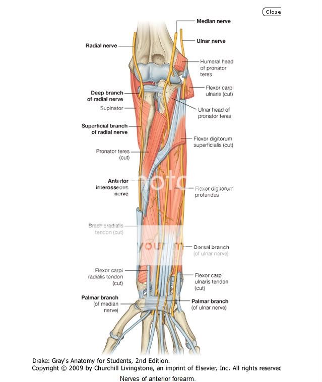
Nerves in the anterior compartment of the forearm are the median and ulnar nerves, and the superficial branch of the radial nerve (Fig. 7.87).

Median nerve

The median nerve innervates the muscles in the anterior compartment of the forearm except for the flexor carpi ulnaris and the medial part of the flexor digitorum profundus (ring and little fingers). It leaves the cubital fossa by passing between the two heads of the pronator teres muscle and passing between the humero-ulnar and radial heads of the flexor digitorum superficialis muscle (Fig. 7.87).

The median nerve continues a straight linear course distally down the forearm in the fascia on the deep surface of the flexor digitorum superficialis muscle. Just proximal to the wrist, it moves around the lateral side of the muscle and becomes more superficial in position, lying between the tendons of the palmaris longus and flexor carpi radialis muscles. It leaves the forearm and enters the palm of the hand by passing through the carpal tunnel deep to the flexor retinaculum.

Most branches to the muscles in the superficial and intermediate layers of the forearm originate medially from the nerve just distal to the elbow joint.
  • The largest branch of the median nerve in the forearm is the anterior interosseous nerve, which originates between the two heads of the pronator teres, passes distally down the forearm with the anterior interosseous artery, innervates the muscles in the deep layer (the flexor pollicis longus, the lateral half of flexor digitorum profundus, and pronator quadratus) and terminates as articular branches to joints of the distal forearm and wrist.
  • A small palmar branch originates from the median nerve in the distal forearm immediately proximal to the flexor retinaculum (Fig. 7.87), passes superficially into the hand and innervates the skin over the base and central palm. This palmar branch is spared in carpal tunnel syndrome because it passes into the hand superficial to the flexor retinaculum of the wrist.

Ulnar nerve

The ulnar nerve passes through the forearm and into the hand, where most of its major branches occur. In the forearm, the ulnar nerve innervates only the flexor carpi ulnaris muscle and the medial part (ring and little fingers) of the flexor digitorum profundus muscle (Fig. 7.87).
Body_ID: P007518
The ulnar nerve enters the anterior compartment of the forearm by passing posteriorly around the medial epicondyle of the humerus and between the humeral and ulnar heads of the flexor carpi ulnaris muscle. After passing down the medial side of the forearm in the plane between the flexor carpi ulnaris and the flexor digitorum profundus muscles, it lies under the lateral lip of the tendon of the flexor carpi ulnaris proximal to the wrist.

The ulnar artery is lateral to the ulnar nerve in the distal two-thirds of the forearm, and both the ulnar artery and nerve enter the hand by passing superficial to the flexor retinaculum and immediately lateral to the pisiform bone (Fig. 7.87).

In the forearm the ulnar nerve gives rise to:
  • muscular branches to the flexor carpi ulnaris and to the medial half of the flexor digitorum profundus that arise soon after the ulnar nerve enters the forearm; and
  • two small cutaneous branches-the palmar branch originates in the middle of the forearm and passes into the hand to supply skin on the medial side of the palm; the larger dorsal branch originates from the ulnar nerve in the distal forearm and passes posteriorly deep to the tendon of the flexor carpi ulnaris and innervates skin on the posteromedial side of the back of the hand and most skin on the posterior surfaces of the medial one and one-half digits.

Radial nerve
The radial nerve bifurcates into deep and superficial branches under the margin of the brachioradialis muscle in the lateral border of the cubital fossa (Fig. 7.87).
  • The deep branch is predominantly motor and passes between the two heads of the supinator muscle to access and supply muscles in the posterior compartment of the forearm.
  • The superficial branch of the radial nerve is sensory. It passes down the anterolateral aspect of the forearm deep to the brachioradialis muscle and in association with the radial artery. Approximately two-thirds of the way down the forearm, the superficial branch of the radial nerve passes laterally and posteriorly around the radial side of the forearm deep to the tendon of the brachioradialis. The nerve continues into the hand where it innervates skin on the posterolateral surface.


From MOORE'S

Nerves of Forearm

The nerves of the forearm are the median, ulnar, and radial. The median nerve is the principal nerve of the anterior (flexorpronator) compartment of the forearm (Figs. 6.57B and 6.69A). Although the radial nerve appears in the cubital region, it soon enters the posterior (extensor-supinator) compartment of the forearm. Besides the cutaneous branches, there are only two nerves of the anterior aspect of the forearm: the median and ulnar nerves. The named nerves of the forearm are illustrated in Figure 6.69 and their origins and courses are described in Table 6.13. The following sections provide additional details and discuss unnamed branches.
MEDIAN NERVE IN FOREARM
The median nerve is the principal nerve of the anterior compartment of the forearm (Figs. 6.69A and 6.70; Table 6.13). It supplies muscular branches directly to the muscles of the superficial and intermediate layers of forearm flexors (except the FCU), and deep muscles (except for the medial [ulnar] half of the FDP) via its branch, the anterior interosseous nerve.
The median nerve has no branches in the arm other than small twigs to the brachial artery. Its major branch in the forearm is the anterior interosseous nerve (Fig. 6.69A, Table 6.13). In addition, the following unnamed branches of the median nerve arise in the forearm:
  • Articular branches. These branches pass to the elbow joint as the median nerve passes it.
  • Muscular branches. The nerve to the pronator teres usually arises at the elbow and enters the lateral border of the muscle. A broad bundle of nerves pierces the superficial flexor group of muscles and innervates the FCR, the palmaris longus, and the FDS.
  • Anterior interosseous nerve. This branch runs distally on the interosseous membrane with the anterior interosseous branch of the ulnar artery. After supplying the deep forearm flexors (except the ulnar part of the FDP, which sends tendons to 4th and 5th fingers), it passes deep to and supplies the pronator quadratus, then ends by sending articular branches to the wrist joint.
  • Palmar cutaneous branch of the median nerve. This branch arises in the forearm, just proximal to the flexor retinaculum, but is distributed to skin of the central part of the palm.
ULNAR NERVE IN FOREARM
Like the median nerve, the ulnar nerve does not give rise to branches during its passage through the arm. In the forearm it supplies only one and a half muscles, the FCU (as it enters the forearm by passing between its two heads of proximal attachment) and the ulnar part of the FDP, which sends tendons to the 4th and 5th digits (Fig. 6.69B, Table 6.13). The ulnar nerve and artery emerge from beneath the FCU tendon and become superficial just proximal to the wrist. They pass superficial to the flexor retinaculum and enter the hand by passing through a groove between the pisiform and the hook of the hamate.
A band of fibrous tissue from the flexor retinaculum bridges the groove to form the small ulnar canal (Guyon canal) (Fig. 6.70B). The branches of the ulnar nerve arising in the forearm include unnamed muscular and articular branches, and cutaneous branches that pass to the hand:
  • Articular branches pass to the elbow joint while the nerve is between the olecranon and the medial epicondyle.

  • Muscular branches supply the FCU and the medial half of the FDP.
  • The palmar and dorsal cutaneous branches arise from the ulnar nerve in the forearm, but their sensory fibers are distributed to the skin of the hand.

RADIAL NERVE IN FOREARM
Unlike the medial and ulnar nerves, the radial nerve serves motor and sensory functions in both the arm and the forearm (but only sensory functions in the hand). However, its sensory and motor fibers are distributed in the forearm by two separate branches, the superficial (sensory or cutaneous) and deep radial/posterior interosseous nerve (motor) (Fig. 6.69C & D, Table 6.13). It divides into these terminal branches as it appears in the cubital fossa, anterior to the lateral epicondyle of the humerus, between the brachialis and the brachioradialis (Fig. 6.64). The two branches immediately part company, the deep branch winding laterally around the radius, piercing the supinator en route to the posterior compartment.
The posterior cutaneous nerve of the forearm arises from the radial nerve in the posterior compartment of the arm, as it runs along the radial groove of the humerus. Thus it reaches the forearm independent of the radial nerve, descending in the subcutaneous tissue of the posterior aspect of the forearm to the wrist, supplying the skin (Fig. 6.69D).
The superficial branch of the radial nerve is also a cutaneous nerve, but it gives rise to articular branches as well. It is distributed to skin on the dorsum of the hand and to a number of joints in the hand, branching soon after it emerges from the overlying brachioradialis and crosses the roof of the anatomical snuff box (Fig. 6.65).
The deep branch of the radial nerve, after it pierces the supinator, runs in the fascial plane between superficial and deep extensor muscles in close proximity to the posterior interosseous artery; it is usually referred to as the posterior interosseous nerve (Figs. 6.64 and 6.69C). It supplies motor innervation to all the muscles with fleshy bellies located entirely in the posterior compartment of the forearm (distal to the lateral epicondyle of the humerus).

LATERAL AND MEDIAL CUTANEOUS NERVES OF FOREARM
The lateral cutaneous nerve of the forearm (lateral antebrachial cutaneous nerve) is the continuation of the musculocutaneous nerve after its motor branches have all been given off to the muscles of the anterior compartment of the arm.
The medial cutaneous nerve of the forearm (medial antebrachial cutaneous nerve) is an independent branch of the medial cord of the brachial plexus. With the posterior cutaneous nerve of the forearm from the radial nerve, each supplying the area of skin indicated by its name, these three nerves provide all the cutaneous innervation of the forearm (Fig. 6.69D). There is no “anterior cutaneous nerve of the forearm.” (Memory device: This is similar to the brachial plexus, which has lateral, medial, and posterior cords but no anterior cord.)
Although the arteries, veins, and nerves of the forearm have been considered separately, it is important to place them into their anatomical context. Except for the superficial veins, which often course independently in the subcutaneous tissue, these neurovascular structures usually exist as components of neurovascular bundles. These bundles are composed of arteries, veins (in the limbs, usually in the form of accompanying veins), and nerves as well as lymphatic vessels, which are usually surrounded by a neurovascular sheath of varying density.

Tuesday, July 28, 2009

Histology of the Skin


Hello all, I've tried to come up with some slides and pictures which can help you with tommorow's practical session. Here are a few compilations of slides which I compiled from some websites across the internet. Most of the explanation are of my own so forgive me if it's not really accurate. Enjoy!

CLICK ON THE PICTURES TO ENLARGE AND TO GET A BETTER VIEW.
(Sorry, the pictures look a little off coz its too big)


THICK SKIN The following picture shows the histology slide for thick skin. Things to note for is the thickened keratinized layer or the stratum corneum.

Here is a close up of that layer ( thick purple layer as shown below)


Here shows another view of the whole layer of thick skin.

thick3.jpg picture by whizz35mb


THIN SKIN


Here is a picture of the thin skin: (Notice the much thinner stratum corneum layer as compared to the thick skin)


COMPARISON OF THICK AND THIN SKIN

Here is a side by side comparison of the thick and thin skin:


This plate shows the structural variation of the epidermis in different parts of the body.

The epidermis in all three areas is keratinized stratified squamous epithelium. The thickness varies from one site to the other. Note the marked thickness of the epidermis in C (palm). The epidermis of palms of the hands and soles of the feet is the thickest in the body. In these regions only four layers of the epidermis are well delineated.


SKIN APPENDAGES

Here are the labelled diagrams you can use for the pratical.


MELANOCYTES AND MELANOMA

Here is what melanocytes would look like:
(Notice the brown area? Those are the due to the production of melanin)


As you can see in this picture, the epidermal layer is much much more thicker in melanoma as compared to the normal skin. (Epidermal layer is the one in darker purple)


A closer view at 20x shows disorganisation of the cells. Not sure how else to describe it.


PSORIASIS

Four distinct pathological alterations characterise this disorder:

Inflammation.
Hyperproliferation of the epidermis
Altered maturation of the epidermis (resulting in scaling)
Vascular alterations (which add to redness).


Histology of psoriasis

  1. Marked hyperkeratosis with parakeratosis (abnormal maturation)

  2. Loss of granular layer

  3. Epidermal acathosis and elongation of rete ridges (reflecting hyperproliferative state)

  4. Vascular dilatation (these vessels are abnormal as well). Generalised inflammation can also be seen, with T-lymphocytes in the dermis and epidermis.

    For more info on this : Visit these websites!
    http://www.dermamed.com/tech_docs/psoriasis/psoriasis_lit.asp http://www.dermnet.com/thumbnailIndex.cfm?moduleID=15&moduleGroupID=462&groupIndex=0&numcols=0


Monday, July 27, 2009

Anatomy of the Skin (Integumentary System)

Hey guys...this is what i have found. I used Saladin, Wheater's and a website for this...i wont label the images nor post them up coz they cannot be uploaded here..so i will post the link for you guys to refer to the images aites?...the images cl..early show both thick and thin skin enjoy your night ppl

1. Draw and label a diagram showing histological features of thick skin
2. Draw and label a diagram showing histological features of thin skin for
comparison.
Answer: refer to website-
connectiondev.lww.com/Products/eroschenko/documents/.../Ch10.pdf


3. List the 4 major cell types in epidermis and state their function. What are the key changes seen in disorders involving melanocytes such as vitiligo & malignant melanoma.
Answer:
Keratinocytes-synthesize keratin by a process known as keratinisation or cornification.Keratinocytes of the stratum basale undergo mitosis and produce new epidermal cells to replace the dead ones that exfoliate (flake off) from the surface.

Melanocytes-synthesize the pigment melanin. They have long branching processes that spread among the basal keratinocytes and continually shed melanin-containing fragments from their tips. The keratinocytes phagocytize these fragments and accumulate melanin granules on the “sunny side” of the nucleus. Like a little parasol, the pigment shields the DNA from ultraviolet radiation. People of all skin colors have about equal numbers of melanocytes. Differences in color result from differences in the rate of melanin synthesis and how clumped or spread out the melanin is in the keratinocytes. In light skin, the melanin is less abundant and is relatively clumped near the nucleus, imparting less colour to the cells.

Merkel cells-relatively few in number, are receptors for the sense of touch. The tactile cell and its dermal nerve fiber are collectively called a tactile (Merkel) disc.

Langerhan’s cell-antigen presenting cells(APC),express a large number of lymphocyte and macrophage surface markers.

Vitiligo-common disease in which symmetrical areas of depigmentation of the skin occur,often on the hands,fingers and face. The disease destroys all the melanocytes in the affected skin and skin becomes glaringly white, the keratinocytes are not affected. It is an autoimmune disease and destroys the melanocytes.

Malignant melanoma-malignant tumour of melanocytes, particularly affecting pale-skinned people who are exposed to excessive UV light.


4. With the help of a diagram, highlight the microscopic features of the major skin appendages.
Answer:
Refer to Saladin 4th Ed integumentary System


5. What is psoriasis? Briefly describe the main histopathological features associated with this disease.
Answer:
The transition of keratinocytes from replicating basal cell,through the keratinocytes of the prickle cell layer(stratum spinosum), to the flattening and degenerating granular layer cells(stratum granulosum) packed with tonofibrils and keratinohyaline granules can be regarded as an efficient sequence culminating in the production of a tough resistant keratin layer on the surface of skin. The normal transit time is 50-60 days but psoriasis causes the transition to only take about 7 days. It isa common skin condition with unknown aetiology but with an element of multifactorial inheritance. The maturation process is so rushed that there is insufficient time for full development of tonofibrils and keratohyaline in the stratum spinosum and the granular layer does not form. Instead of normal keratin, there is a surface opaque white scale composed of a mixture of keratin and nuclear and cytoplasmic debris of keratinocytes. The rapidly proliferating epidermis is thickened to produce raised red patches under the white scale.


Hope it helps...XD....

Thursday, July 23, 2009

Additional info for Breasts

Hey guys..here are some additional info from dr kelvins lect..XD
Nerves supplying serratus anterior
-long thoracic nerve( if cut leads to winging of scapula)
-thoracodorsal neurovascular bundle9latissimus dorsi nerve)

Seroma-collection of fluid after mastectomy

SEntinel Lymph Node Biopsy- inject dye( isosulphan blue)
-Detect 1st node the dye travels to.
-If infected then very high chance of metastasis.
-If not infected, lower chance of metastasis.

Anatomy of the Breast

Anatomy of the Breast.

I've taken the answers from Moore's, Gray's Antomy and also Saddler's Embryology

1.1 What is the embryological explanation of accessory nipples?

The first indication of mammary glands is found in the form of a band-like thickening of the epidermis, the mammary line or mammary ridge. In a 7-week embryo, this line extends on each side of the body from the base of the forelimb to the region of the hindlimb. (Although the major part of the mammary line disappears shortly after it forms, a small portion in the thoracic region persists and penetrates the underlying mesenchyme . Here it forms 16 to 24 sprouts, which in turn give rise to small, solid buds. By the end of prenatal life, the epithelial sprouts are canalized and form the lactiferous ducts, and the buds form small ducts and alveoli of the gland. Initially, the lactiferous ducts open into a small epithelial pit. Shortly after birth, this pit is transformed into the nipple by proliferation of the underlying mesenchyme.

Polythelia is a condition where accessory nipples have formed due to the persistence of fragments of the mammary line. Accessory nipples may develop anywhere along the original mammary line, but usually appear in the axillary region.

Polymastia occurs when a remnant of the mammary line develops into a complete breast.

2.1 What is the arterial supply of the breast?

The arterial supply of the breast derives from the:

  • Medial mammary branches of perforating branches and anterior intercostal branches of the internal thoracic artery, originating from the subclavian artery.
  • Lateral thoracic and thoracoacromial arteries, branches of the axillary artery.
  • Posterior intercostal arteries, branches of the thoracic aorta in the 2nd, 3rd, and 4th intercostal spaces.

3.1 How may Cooper’s ligaments be evident in breast carcinoma?

Interference with the lymphatic drainage by cancer may cause lymphedema (edema, excess fluid in the subcutaneous tissue), which in turn may result in deviation of the nipple and a thickened, leather-like appearance of the skin. Prominent or “puffy” skin between dimpled pores give it an orange-peel appearance (peau d'orange sign). Larger dimples (fingertip size or bigger) result from cancerous invasion of the glandular tissue and fibrosis (fibrous degeneration), which causes shortening or places traction on the suspensory ligaments. Subareolar breast cancer may cause retraction of the nipple by a similar mechanism involving the lactiferous ducts.

A well-developed, connective tissue stroma surrounds the ducts and lobules of the mammary gland. In certain regions, this condenses to form well-defined ligaments, the suspensory ligaments of breast, which are continuous with the dermis of the skin and support the breast. Carcinoma of the breast creates tension on these ligaments, causing pitting of the skin. Subcutaneous lymphatic obstruction and tumor growth pull on connective tissue ligaments in the breast resulting in;ll the appearance of an orange peel texture (peau d'orange) on the surface of the breast. Further subcutaneous spread can induce a rare manifestation of breast cancer that produces a hard, woody texture to the skin (cancer en cuirasse).

3.2 How may carcinoma spread via the lymphatics/veins/local invasion?

Breast cancer typically spreads by means of lymphatic vessels (lymphogenic metastasis), which carry cancer cells from the breast to the lymph nodes, chiefly those in the axilla. The cells lodge in the nodes, producing nests of tumor cells (metastases). Abundant communications among lymphatic pathways and among axillary, cervical, and parasternal nodes may also cause metastases from the breast to develop in the supraclavicular lymph nodes, the opposite breast, or the abdomen. Because most of lymphatic drainage of the breast is to the axillary lymph nodes (Fig. 1.24A), they are the most common site of metastasis from a breast cancer. Because most of lymphatic drainage of the breast is to the axillary lymph nodes (Fig. 1.24A), they are the most common site of metastasis from a breast cancer. Enlargement of these palpable nodes suggests the possibility of breast cancer and may be key to early detection. However, the absence of enlarged axillary lymph nodes is no guarantee that metastasis from a breast cancer has not occurred because the malignant cells may have passed to other nodes, such as the infraclavicular and supraclavicular lymph nodes.

Carcinoma of the breast usually arises in the upper outer quadrant and first spreads to the axillary nodes. However, medial breast lesions may drain through the chest wall to the nodes along the internal mammary artery. Thereafter, in both instances, the supraclavicular and infraclavicular nodes may be seeded. In some cases, the cancer cells seem to traverse the lymphatic channels within the immediately proximate nodes to be trapped in subsequent lymph nodes, producing so-calledskip metastases. The cells may traverse all of the lymph nodes ultimately to reach the vascular compartment via the thoracic duct.

4.1 How will you palpate the axillary lymph nodes?
Refer to PDF file. http://qap.sdsu.edu/resources/tools/pdf/lymphnode.pdfv

5.1 Describe a normal mammogram

No thickening of the skin, no calcification and no dominant masses.

Radiographic examination of the breasts, mammography, is one of the techniques used to detect breast masses. A carcinoma appears as a large, jagged density in the mammogram. The skin is thickened over the tumor (upper two arrows in) and the nipple is depressed. Surgeons use mammography as a guide when removing breast tumors, cysts, and abscesses.